Imaging and Surveillance of Chronic Aortic Dissection
Updated: February 17, 2022
- This scientific statement considers measurement techniques for patients with chronic aortic dissection, including the need for standardized measurements in life-long surveillance. It examines the role of imaging and computer simulations to predict aortic false lumen degeneration, remodeling, and biomechanical failure from morphologic and hemodynamic features that could improve risk stratification and individualize treatment options to support patients.
- Patients surviving an acute aortic dissection require continued life-long surveillance of their diseased aorta. Late complications, as seen in chronic false lumen degeneration and aneurysm formation, often require surgical, endovascular or hybrid interventions to treat or prevent aortic rupture.
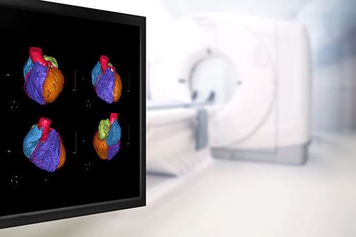
- Imaging plays a central role in the surveillance of patients with chronic aortic dissection. Monitoring the inevitable degeneration and aneurysmal dilatation of residual aortic false lumen is critical for medical decisions and timing of interventions.
Supporting Materials
- Commentary: Is Imaging the Holy Grail of Aftercare in Aortic Dissection? by Christoph A. Nienaber MD, PhD (1,2); Fortunate Rusike BSc (1); Xun Yuan MBBS, MMED (1,2)
- Top Things to Know: Imaging & Surveillance of Chronic Aortic Dissection